
Click here to see all images
May, 2024
Case of the Month
Clinical History:
A middle-aged non-smoking woman with history of untreated asthma upon exertion underwent a CT scan for a respiratory tract infection, which led to the incidental discovery of a 10mm solid lung nodule in the left upper lobe. The lesion was stable on follow-up and was removed in a segmentectomy 2 years later upon patient request.
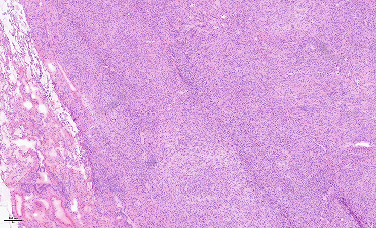
Macroscopically, the nodule was well-circumscribed, tan-white with a homogenous consistency. An intraoperative frozen section was performed (Figure 1 and 2). Representative histological images (Figure 3-5) and immunohistochemical stains are provided (Figure 6-8).
Q1. What is the most likely diagnosis
- Sclerosing pneumocytoma
- Pleomorphic carcinoma of the lung
- Carcinoid tumor of the lung
- Pulmonary hamartoma
Q2. Which of the following immunohistochemical markers is most likely to be strongly positive in both neoplastic cell populations?
- CK7
- EMA
- Synaptophysin
- ER
Q3. Which genetic alteration is most likely to be found in this lesion?
- KRAS
- MEN1
- AKT1
- EGFR
Answers to Quiz
Q2. B
Q3. C
Diagnosis
Discussion
Sclerosing pneumocytoma is a benign pulmonary neoplasm with an unknown pathogenesis, presumably deriving from primitive pulmonary epithelium. Nearly all sclerosing pneumocytomas harbor AKT1 alterations, which can be useful for diagnosis confirmation. The essential diagnostic criteria according to WHO 2021 are the presence of a well-circumscribed lesion composed of a dual population of cuboidal surface cells and stromal round cells, usually featuring a mixture of papillary, sclerotic, solid, and hemorrhagic growth patterns. The distinctive immunostaining profile of TTF1-and EMA-positivity in both surface and round cells and absence or weak staining of cytokeratin in the round cells, while the surface cells are strongly positive, is desirable.
The clinical course is typically indolent, with most lesions being discovered incidentally, as in the current case. However, cases with recurrence, malignant transformation and lymph node and distant metastases have been described. Rarely sclerosing pneumocytoma may co-exist with carcinoid tumors or adenocarcinoma.
Treatment consists of surgical resection, which is usually curative.
Take home message for trainees:
The presence of a well-circumscribed tumor with a dual cell population in a non-smoker should raise the suspicion for sclerosing pneumocytoma. Significant cell atypia can present a diagnostic pitfall, especially on intraoperative frozen section.
References
Miyagawa-Hayashino A, Tazelaar HD, Langel DJ, Colby TV. Pulmonary sclerosing hemangioma with lymph node metastases: report of 4 cases. Arch Pathol Lab Med. 2003 Mar;127(3):321-5.
Wang X, Ng CS, Shi X, Yin W. Characteristics of metastatic and nonmetastatic pulmonary sclerosing pneumocytomas: a clinicopathological study of 68 cases and 15 reported metastatic cases. Lab Invest. 2023 Jul;103(7):100135.
Yeh YC, Chu PY, Lin SY, et al. Comprehensive genomic and transcriptomic analysis of sclerosing pneumocytoma. Mod Pathol. 2024 Jan;37(1):100354.
Contributors
Resident
Institute of Pathology
Lausanne University Hospital and University of Lausanne
Lausanne, Switzerland
Sabina Berezowska, MD
Associate Professor
Institute of Pathology
Lausanne University Hospital and University of Lausanne
Lausanne, Switzerland